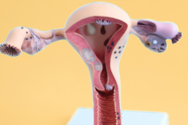
Adenomyosis, how does it affect pregnancy?
At Clinica Fertia we are committed and work to be able to help our patients in cases that are considered highly complex in gynaecology. One of these cases is adenomyosis.
Adenomyosis and its evolution throughout history
The first detailed pathological description of endometriosis and adenomyosis was published in Vienna in 1860 by Karl Freiherr Von Rokitansky.

Adenomyosis is a benign uterine disease defined as the presence of heterotopic endometrial glands and stroma in the myometrium with adjacent smooth muscle hyperplasia. It is a common benign uterine disorder and has been considered a histopathological diagnosis made after hysterectomy, classically performed in perimenopausal women with abnormal uterine bleeding (SUA) or pelvic pain with a prevalence of 30%. Until recently, adenomyosis was a clinically neglected disease. Nowadays, adenomyosis can also be diagnosed using non-invasive techniques, thanks to advances in imaging, such as MRI and transvaginal ultrasound. Thus, a new epidemiological scenario has developed and adenomyosis has become a multifaceted disease diagnosed by imaging techniques in young women with abnormal uterine bleeding, infertility or pelvic pain, even in asymptomatic women. It is often diagnosed in association with other gynaecological comorbidities such as endometriosis and uterine fibroids.
Is there a relationship between adenomyosis and infertility?
Several studies have shown a relationship between adenomyosis and infertility, but the precise mechanisms involved are not clearly established. Eutopic and ectopic endometrial tissues from patients with adenomyosis show a different expression of genes associated with apoptosis and angiogenesis such as Bcl-2 and VEGF (1), there is a higher expression of VEGF involved in angiogenic processes as well as in vascular permeability, increased expression of Bcl-2, an anti-apoptotic molecule, present throughout the secretory phase, this overexpression increases cellular resistance to apoptosis, which could influence endometrial tissue remodelling during blastocyst implantation and placental development (2). Other factors involved in implantation appear to be altered, such as increased prostaglandin levels in the ectopic endometrial epithelium, increased expression of cytochrome P450 aromatase in the eutopic endometrium, decreased integrin beta 3, osteopontin and leukemia inhibitory factor, and reduced HOXA-10 gene function during the implantation window. Decreased expression of progesterone A and B receptors has also been described in ectopic endometrium, probably related to epigenetic changes. This resistance to progesterone could lead to abnormal expression of progesterone receptor-related genes and reduced expression of implantation-related genes (3). In addition to these changes in endometrial implantation-related genes, women with adenomyosis have reduced uterotubular transport, increased levels of nitric oxide in the uterine cavity, altered contractility, and altered uterine cavity volume, among other factors, with a significant negative impact on fertility.
What was the impact of adenomyosis on achieving pregnancy and pregnancy complications?
Six recent meta-analyses have assessed the impact of adenomyosis on pregnancy complications, as well as fertility outcome after assisted reproduction and natural conception.
The first, conducted by Maheshwari in 2012 (4), established the need for accurate diagnosis as, given the delay in pregnancy and improved diagnosis, the likelihood of diagnosing adenomyosis in the infertile population increases. Vercellini in 2014 (5) found that women with adenomyosis had a 28% reduction in the likelihood of clinical pregnancy and an increased risk of early termination of pregnancy, so screening for adenomyosis should be encouraged before attempting assisted reproductive procedures. Margit Dueholm in 2017 (6) also found a reduction in implantation, early pregnancy loss and premature birth related to adenomyosis, in her meta-analysis some studies indicate an adverse implantation outcome related to the magnitude of change in JZ ( 7). Studies that assessed the morphology of the endometrial cavity showed moderate distortion in 23% of women and 10% were severely impacted with a pseudo T-shaped uterus (8). In 2017, Younes G (9) published a meta-analysis of 11 different studies in which it appears that diffuse adenomyosis is worse than focal or localised adenomyosis. They also found that women with adenomyosis had a higher miscarriage rate than those without adenomyosis, the presence of adenomyosis was associated with a 41% decrease in live birth rate. They conclude that adenomyosis has a detrimental effect on the clinical outcome of in vitro fertilisation. Subsequently, Horton’s group (10) published another meta-analysis in 2019 that showed a reduction in clinical pregnancy rate, a reduction in live birth rate and an increase in miscarriage rate in women with adenomyosis compared to controls. A lower implantation rate after in vitro fertilisation, although there is no difference in oocyte yield. An increased risk of late pregnancy and neonatal complications such as premature birth, placenta previa, pregnancy-induced hypertension, postpartum haemorrhage.
The most recent meta-analysis published in 2021 by Konstantinos Nirgianakis (1), finds that adenomyosis is significantly associated with a lower pregnancy rate after treatment with assisted reproductive technology, a higher rate of miscarriages. It is also significantly associated with an increased risk of pre-eclampsia, premature birth, caesarean section, incorrect fe
What type of specific therapy in assisted reproduction is most appropriate in women with adenomyosis?
Currently, pre-treatment with Gn-Rh-a is suggested before natural conception in women without reduced ovarian reserve. Gn-Rh agonists transiently suppress the hypothalamic-pituitary-ovarian axis and induce a hypoestrogenic effect, resulting in a reduction in adenomyosis. It has also been shown that Gn-Rh agonists can exert antiproliferative and apoptotic effects in cultures of endometriosis cells and in some tumour cells derived from reproductive organs (12). Treatment with Gn-Rh agonists significantly suppressed the proliferation of cells derived from the endometrium and pathological lesions in patients with adenomyosis. There are no randomised controlled trials comparing different in vitro fertilisation protocols in women with adenomyosis. Although many authors report a higher rate of pregnancy and live births by adopting an ultra-long Gn-Rh -a protocol. The use of the Gn-Rh agonist induces apoptosis and reduces inflammatory reaction and angiogenesis. The disadvantages of long-term use of Gn-Rh are longer ovarian stimulation, higher gonadotropin doses and lower oocyte yield.
Therefore, its use is reasonable in patients with normal ovarian reserve, especially in patients with diffuse adenomyosis. Other authors believe that the use of the GnRh analogue is more profitable before frozen embryo transfer, especially in women with low ovarian reserve. In Niu’s study (13), frozen embryo transfers in women with adenomyosis, following the use of a long-term GnRh agonist plus hormone replacement therapy, had a significantly higher clinical pregnancy rate, implantation rate and ongoing pregnancy than the HRT group without prior GnRH treatment.
Other treatment options such as pretreatment prior to frozen embryo transfer in women with adenomyosis, use of levonorgestrel-releasing intrauterine system three months prior, conventional FET cycle also showed higher implantation rate, clinical pregnancy rate and lower miscarriage rate than patients with adenomyosis and conventional FET cycle (14).
All of which leads us to conclude that…
It can be concluded that adenomyosis is associated with adverse effects on fertility after ART and, irrespective of the mode of conception, adenomyosis is also associated with adverse outcomes in pregnancy and the newborn. There is an urgent need for adequate preconception counselling and careful monitoring of pregnancy in patients with adenomyosis. The gynaecologist needs to be aware of these risks in order to establish adequate pregnancy monitoring in these patients to allow early diagnosis and treatment of pregnancy complications. Personalised antiretroviral therapies should be applied to these patients to improve their chances of conception.
Gynaecological check-ups are a very important factor in the detection of this disease and its resolution from the first moment it is detected. There is no set age for visiting a gynaecologist, but for women a routine check-up every year is very important. At Clinica Fertia we insist on control and health of our patients.

Written by:
Dr. Elena Puente
Medical director of Clinica Fertia
Bibliografía
- Yalaza C,CanacanKatan N, Gurses I, Tasdelen B.Altered VEGF, Bcl-2 and IDH1 expression in patients with adenomyosis.Archives of Gynecol and Osbtet.2020. 2. Vatansever HS,Lacin S,Ozbilgin MK.Changed Bcl:bax ratio in endometrium of patients with unexplained infertility. Acta Histchem.2005;107.345-355. 3. Mehasseb MK,Panchal R, Taylor AH. Estrogen and progesterone receptor isoform distribution through the menstrual cycle in uteri with and without adenomyosis.Fertil Steril.2009;92:886-889. 4. Maheshwari A, Gurunath S, Fatima F, Bhattacharya S. Adenomyosis and subfertility: a systematic review of prevalence, diagnosis, treatment and fertility outcomes. Hum Reprod Update.2012;18:374-392. 5.Vercellini P, Consoni D, Dridi D,Bracco B, Frattaruolo MP, Somiglianna E. Uterinne adenomyosis and in vitro fertilization outcome:a systematic review and meta-analysis.Hum Reprod 2014b.29:964-977. 6. Dueholm M. Uterine adenomyosis and infertility, review of reproductive outcome after in vitro and surgery.Acta Obstet Gynecol Scand.2017;96:715-726. 7. Maubon A, Faury A, Kapella M, Pouquet M, Piver P. Uterine junctional zonne at magnetic resonance imaging:a predictor of in vitro fertilization failure.J Obstet Gynecol res.2010:36:611-618. 8.. Puente JM, Fabris A, Patel J, Patel A,Cerrillo M, Requena A.Adenomyosis in infertile women:prevalence and the role of 3D ultrasound as a marker of severity of the disease.Reprod Biol Endocrinol.2016;14:60. 9. Younes G, Tulandi T.Effects of adenomyosis on in vitro fertilization outcomes: a meta-analysis.Fertil Steril.2017;108:483-490.) 10. Horton J, Sterrenburg M, Lane S, Maheshwari A, li TC, Cheong Y. Reproductive , obstetric, and perinatal outcomes of women with adenomyosis and endometriosis: A systematic review and meta-analysis.Hum Reprod update.2019;25:592-632. 11. Nirgianakis K, Kalaitzopoulos DR, Scharwtz AS, Spaanderman M, Kramer BW, Mueller MD, Mueeller M. Fertility, pregnancy and neonatal outcomes of patients with adenomyosis: a systematic review and meta-analysis. Reprod Biom Online.2021: 42: 185-206. 12. Khan KN, Kitajima M, Hiraki K. Cell proliferation effect of Gn-Rh agonist on pathological lesions of women with endometriosis,adenomyosis and uterine myoma. Hum reprod.2010;25:2878-2890). 13. Niu Z, Chen Q, Sun Y,Feng Y. Long term pituitary downregulation before frozen embryo transfer could improve pregnancy outcome in women with adenomyosis.Gynecol Endocrinol.2013;29:1026-1030. 14. Liang Z, Yin M, Wang Y, Kuang Y. Effect of pretreatment with a levonorgestrel-releasing intrauterine system on IVF and vitrified -warmed embryo transfer outcomes in women with adenomyosis.Reprod Biomed Online.2019;39:111-118.
